
Welcome to the Brachiopod subsite on Invertebrate Paleontology!
Please enter a genera name to retrieve more information.
Diholkorhynchia
Classification
Phylum:
Brachiopoda
Subphylum:
Rhynchonelliformea
Class:
Rhynchonellata
Order:
Rhynchonellida
Superfamily:
Norelloidea
Family:
Norellidae
Subfamily:
Diholkorhynchiinae
Formal Genus Name and Reference:
Diholkorhynchia Y ANG & X U , 1966, p. 24[99]
Type Species:
Rhynchonella sinensis KOKEN, 1900, p. 206, OD
Images
(Click to enlarge in a new window)
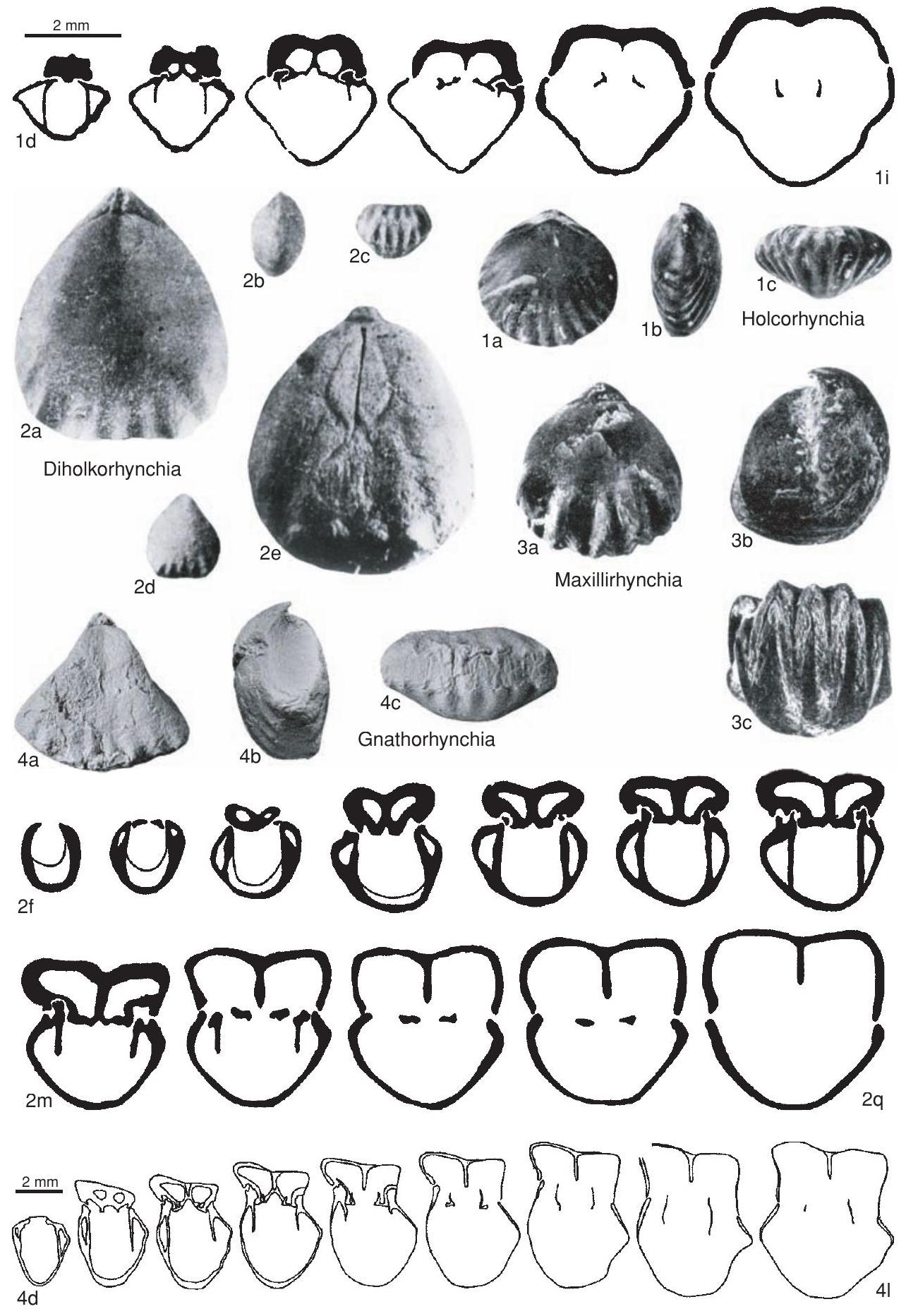 Fig. 892, 2a-q. *D. sinensis (Koken), Anisian, Gheizhou, a, syntype, dorsal view, MCMB DDM4, x3, b-d, syntype, lateral, anterior, ventral views, MCMB DDM4, x1, e, syntype, dorsal view of steinkern with muscle scars, ×3; f–q, paratype, transverse serial sections, distances in mm from ventral umbo, 0.4, 0.8, 1.0, 1.3, 1.5, 1.6, 1.8, 2.0, 2.2, 2.4, 2.6, 2.9, MCMB DDKC 120 *4-2 (Yang & Xu, 1966).
Synonyms
Dihorhynchia
Geographic Distribution
southwestern China
Age Range
Beginning Stage in Treatise Usage:
Middle Triassic
Beginning International Stage:
Anisian
Fraction Up In Beginning Stage:
0
Beginning Date:
246.7
Ending Stage in Treatise Usage:
Middle Triassic
Ending International Stage:
Ladinian
Fraction Up In Ending Stage:
100
Ending Date:
237
Description
Small, triangular to subpentagonal, with short hinge, biconvex, anterior commissure multiplicate, beak small, straight or strongly incurved, pedicle opening small, oval, deltidial plates conjunct, ventral sulcus well developed and limited to anterior half of shell, dorsal valve regularly convex, but with medial depression starting near umbonal area, widening at slightly anterior of middle of dorsal length, and weakening on fold, shell completely smooth posteriorly and marked only anteriorly and laterally with short plicae, costellae absent. Dental plates almost parallel, muscle scars pear shaped, pallial markings bifurcated in ventral interior. Dorsal interior with well-developed hinge plates, septalium, median septum, muscle scars oval, situated at both sides of median septum, pallial markings bifurcated.
References
Museum or Author Information
Classification
Phylum:
Brachiopoda
Subphylum:
Rhynchonelliformea
Class:
Rhynchonellata
Order:
Rhynchonellida
Superfamily:
Norelloidea
Family:
Norellidae
Subfamily:
Diholkorhynchiinae
Formal Genus Name and Reference:
Diholkorhynchia Y ANG & X U , 1966, p. 24[99]
Type Species:
Rhynchonella sinensis KOKEN, 1900, p. 206, OD
Images
(Click to enlarge in a new window)
Fig. 892, 2a-q. *D. sinensis (Koken), Anisian, Gheizhou, a, syntype, dorsal view, MCMB DDM4, x3, b-d, syntype, lateral, anterior, ventral views, MCMB DDM4, x1, e, syntype, dorsal view of steinkern with muscle scars, ×3; f–q, paratype, transverse serial sections, distances in mm from ventral umbo, 0.4, 0.8, 1.0, 1.3, 1.5, 1.6, 1.8, 2.0, 2.2, 2.4, 2.6, 2.9, MCMB DDKC 120 *4-2 (Yang & Xu, 1966).
Synonyms
Dihorhynchia
Geographic Distribution
southwestern China
Age Range
Beginning Stage in Treatise Usage:
Middle Triassic
Beginning International Stage:
Anisian
Fraction Up In Beginning Stage:
0
Beginning Date:
246.7
Ending Stage in Treatise Usage:
Middle Triassic
Ending International Stage:
Ladinian
Fraction Up In Ending Stage:
100
Ending Date:
237
Description
Small, triangular to subpentagonal, with short hinge, biconvex, anterior commissure multiplicate, beak small, straight or strongly incurved, pedicle opening small, oval, deltidial plates conjunct, ventral sulcus well developed and limited to anterior half of shell, dorsal valve regularly convex, but with medial depression starting near umbonal area, widening at slightly anterior of middle of dorsal length, and weakening on fold, shell completely smooth posteriorly and marked only anteriorly and laterally with short plicae, costellae absent. Dental plates almost parallel, muscle scars pear shaped, pallial markings bifurcated in ventral interior. Dorsal interior with well-developed hinge plates, septalium, median septum, muscle scars oval, situated at both sides of median septum, pallial markings bifurcated.